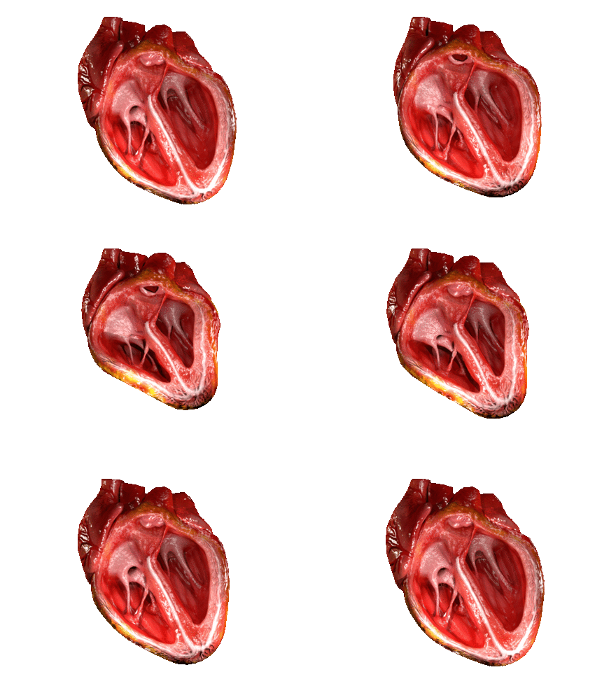
How does the heart work?
The heart is a large muscle that is divided into a right and left side by an inner wall called the septum. The right side of the heart receives deoxygenated blood from the body and then pumps the blood to the lungs to receive fresh oxygen. The left side of the heart receives the oxygenated blood from the lungs and then pumps the blood out the aorta to the body. To review the anatomy of the heart see: What are the parts of the heart. and to review the path blood takes through the heart and body see: What is the path blood takes through the circulatory system.

Computer Simulation of Beating Heart: Dr JanaOfficial
What allows the heart to pump blood through the body?
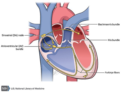
The contraction of heart muscle is initiated by electrical impulses known as action potentials. The rate at which these impulses fire controls the rate of cardiac contraction (the heart rate). The cells that create these rhythmic impulses, setting the pace for blood pumping, are called pacemaker cells, and they directly control the heart rate. These cells are referred to as the cardiac pacemaker which is the natural pacemaker of the heart. In most humans, the concentration of pacemaker cells is located in the sinoatrial (SA) node (see image below).

What is the electrical conduction system of the heart?
The sinoatrial node (SA node) is responsible for the initiation of the cardiac cycle (heartbeat). It spontaneously generates an electrical impulse, which after conducting throughout the heart causes the heart to contract. Although the electrical impulses are generated spontaneously, the rate of the impulses (and therefore the heart rate) is set by the nerves innervating the sinoatrial node. The sinoatrial node is located in the myocardial wall near where the sinus venarum (posterior part of the right atrium) joins the right atrium (upper chamber); hence sino- + atrial. Although some of the heart's cells have the ability to generate the electrical impulses (or action potentials) that trigger cardiac contraction, the sinoatrial node normally initiates it, simply because it generates impulses slightly faster than the other areas with pacemaker potential.
Electrical signals arising in the SA node stimulate the atria to contract and travel to the atrioventricular node (AV node). The AV node is an area of specialized tissue between the atria and the ventricles of the heart. After a delay, the stimulus diverges and is conducted through the left and right bundle of His to the respective Purkinje fibers for each side of the heart, as well as to the endocardium at the apex of the heart, then finally to the ventricular epicardium.
For the heart to maximize efficiency there must be sustantial atrial to ventricular delay to allow the atria to completely empty their contents into the ventricles. Simultaneous contraction would cause inefficient filling and backflow. The ventricles must maximize systolic pressure to force blood through the circulation, so all the ventricular cells must work together.
Cardiac arrhythmias can cause heart block, in which the contractions lose any useful rhythm. In humans, and occasionally in animals, a mechanical device called an artificial pacemaker (or simply "pacemaker") may be used after damage to the body's intrinsic conduction system to produce these impulses synthetically.
What is an Electrocardiogram?
Electrocardiography (ECG or EKG*) is the process of recording the electrical activity of the heart over a period of time using electrodes placed on the skin. These electrodes detect the tiny electrical changes on the skin that arise from the heart muscle's electrophysiologic pattern of depolarizing during each heartbeat. It is a very commonly performed cardiology test.
Test your Understanding:
Related Sites
- Why is carbon monoxide so dangerous?
- The Hemoglobin S molecule and Sickle Cell Anemia
- What is the human genome and how big is it?
- How are living things classified?
- What is the path blood takes through the circulatory system?
- Where is food digested?
- Gifted and Talented STEM
- EDinformatics Science Challenge